What are the differential diagnoses for chordoma?
What are the differential diagnoses for chordoma? Chordoma is a rare type of cancer that grows in the bones of the spine and base of the skull. When doctors look for what might cause symptoms, they think about other conditions too, not just chordoma. It’s important to know all possible causes because treatment can be very different. A good diagnosis helps patients get the right care quickly.Doctors often see similar signs in several medical conditions which can make finding the correct illness tricky. For diseases like chordoma symptoms may show up slowly over time and be quite mild at first. Patients need doctors who pay close attention and consider all possibilities to avoid missing anything.
Finding out if someone has chordoma involves looking at symptoms carefully and using special medical tests. Other illnesses can have overlapping features with this condition so it takes skill to tell them apart. Each patient gets a unique plan based on their test results and how they feel. Quick action after getting a diagnosis leads to better chances of managing health well over time.
Symptoms of Chordoma
Chordoma often starts without any clear signs which can make early diagnosis tough. As it grows one might feel pain in the back or neck area. Sometimes people with chordoma get headaches that don’t seem to go away. This happens because the tumor presses on nerves and other structures. Knowing these symptoms is key for doctors to suspect chordoma.
This medical condition may also lead to weakness or numbness in arms or legs. If the tumor is near the base of the skull it could affect how well you swallow. Some patients notice changes in their voice or even facial expression due to muscle impacts. These are clues that signal doctors towards a potential diagnosis of chordoma.
Another common symptom linked with this illness involves trouble when going to the bathroom. It’s not unusual for individuals with lower spine tumors to have bowel or bladder issues. Recognizing this link helps healthcare providers narrow down differential diagnoses and focus on conditions like chordoma.
Imaging Tests
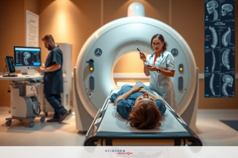
Imaging tests are crucial in the diagnosis process of chordoma. They let doctors see inside the body without surgery. X-rays can show if bones where chordoma occurs have changes. But they’re just a first step because x-rays alone don’t give all the details needed. More advanced imaging is often necessary to confirm chordoma.
Magnetic resonance imaging, or MRI, provides clear pictures of soft tissues and bones affected by chordoma. It uses powerful magnets and radio waves to create detailed images. With MRI doctors can spot tumors in spine areas that other tests might miss. This helps them make accurate differential diagnoses when considering conditions similar to chordoma.
Another important test is computed tomography commonly known as a CT scan. CT scans use special x-ray equipment to get images from different angles around the body. These pictures come together to form a cross-sectional view of internal structures which aids in detecting abnormalities related to chordoma.
Biopsy Procedure
A biopsy is a key step in confirming a diagnosis of chordoma. In this procedure doctors take a small piece of

tissue from the suspected area. It’s done to look at cells under a microscope and see if they are cancerous. For people with symptoms pointing toward chordoma a biopsy provides clear evidence. This test helps rule out other conditions that might seem similar.
The process starts by pinpointing the exact location for sampling often guided by imaging tests already discussed. Then using local anesthesia to numb the spot ensures patient comfort during tissue removal. The doctor uses special tools to get the sample without affecting surrounding areas much. Afterward there might be some soreness but it usually goes away quickly.
Once taken the tissue sample goes straight to a lab where experts check it for signs of chordoma or other medical conditions. They use stains and markers which help them spot unique cell features found in this type of cancer specifically. Results from this analysis give patients and their doctors vital info that guides further treatment choices accurately.
Treatment Options
Once chordoma is diagnosed treatment planning can begin. Surgery is often the first option considered by medical teams. The goal of surgery is to remove as much of the tumor as possible without harming vital functions. Skilled surgeons work carefully around critical structures like nerves and blood vessels. Post- surgery regular check-ups are necessary to monitor for any signs of recurrence.
Radiation therapy may follow surgery or be used when surgical removal isn’t possible. It involves targeting the tumor with high-energy beams to destroy cancer cells left behind. Precision techniques like proton beam therapy minimize damage to nearby healthy tissues while focusing on chordoma areas.
In some situations doctors might suggest a combination of surgery and radiation therapy for better outcomes. Combining treatments aims at reducing the chances that chordoma will come back after initial management efforts are complete. Patients undergoing combined methods usually have close follow-up care plans set up by their healthcare team.
Frequently Asked Questions
What is chordoma?
Chordoma is a rare type of cancer that occurs in the bones of the spine and skull base.
How is chordoma diagnosed?
Diagnosis typically involves imaging tests like MRI or CT scans followed by a biopsy to examine tissue for cancer cells.
What are the treatment options for chordoma?
Treatment may include surgery, radiation therapy, and in some cases, chemotherapy or targeted therapies. The specific plan depends on each patient's situation.


